Prostate cancer care solutions
A clear path for prostate cancer care
Bring the power of MR imaging into your urology practice to support you at every step of a prostate cancer patient’s journey. This allows you to confidently deliver the precise care your patients deserve. Our prostate cancer care solutions features DynaCAD Prostate for advanced visualization and analysis of multi-parametric MR imaging, UroNav for live MR/ultrasound fusion-guided biopsy and therapy procedures, and DynaCAD Urology for urology data management.

Advanced, intuitive image guidance can illuminate the way to a clear path for prostate cancer care.
22 of the top 25 cancer hospitals in the US* use our MRI/Ultrasound fusion prostate cancer solution to support diagnosis.
Featured products in prostate cancer care
-
UroNav
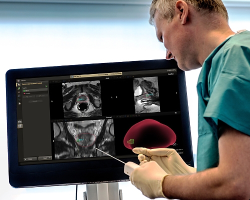
UroNav fuses pre-biopsy MR images of the prostate with ultrasound-guided biopsy images in real time, for excellent delineation of the prostate and suspicious lesions, as well as clear visualization of the biopsy needle path. Combining electromagnetic tracking and navigation with an onboard computer and a real-time imaging interface, UroNav brings precision targeting to your clinical practice in one easy-to-use, mobile workstation.
784026 -
DynaCAD Prostate
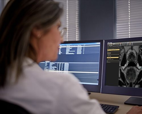
Philips DynaCAD Prostate is an advanced visualization system that empowers you with a comprehensive set of tools for real-time analysis, review, and reporting of multi-parametric MRI studies. Create time and workflow efficiency by transferring images directly from the MRI to DynaCAD. Utilize its robust, automatic post-processing tools and display results in customized hanging protocols for analysis and reporting. At case completion, you can automatically transfer key images, statistical data, and prostate PI-RADS® reports to PACS for archiving. By setting everything up for you to work, DynaCAD helps you enhance your confidence and productivity – so patients get the prompt, precise care they need.
784029 -
DynaCAD Urology
DynaCAD Urology is a purpose build solution that empowers urologists with a dedicated set of tools for utilizing multi-parametric MR data in fusion biopsy workflows. It also provides a solution for managing patients’ biopsy data in urology.
784044


- 0

Fuse diagnostic MR images with live ultrasound for guided procedures and clear delineation of the prostate and lesions.
DynaCAD Prostate offers direct image transfer from your MR scanner for time and workflow efficiency. Analysis and reporting of mpMRI studies are made easy thanks to its robust, automatic post‑processing tools and the ability to display results in customized hanging protocols. At case completion, key images, statistical data and prostate PI-RADS® reports can be automatically transferred to PACS for archiving. DynaCAD Prostate helps you enhance confidence and productivity for precise patient care.
Gain a comprehensive understanding of your urology patient’s progress
DynaCAD Urology provides urologists with an easy way to store, review and manage comprehensive diagnostic and therapeutic data. Create time and workflow efficiencies in your prostate cancer care program. The device allows you to build on the work radiology has done and gives you easy access to your own longitudinal data for case management. By connecting directly to the UroNav system, it also documents your patient’s biopsies and focal treatments.
Perform prostate biopsies with imaging guidance
UroNav is designed to support you with image guidance during prostate biopsies and focal therapy procedures. It fuses diagnostic MR images of the prostate with live ultrasound images for guided procedures in real time. You get clear delineation of the prostate and suspicious lesions, as well as clear visualization of the needle path. Combine UroNav with DynaCAD Urology for a comprehensive solution that supports you at every step of the prostate cancer patient journey.


Footnotes
Products not available for sale in all countries. Please contact your sales representative to ascertain availability in your country.
[1] DynaCAD Urology and DynaCAD Prostate are modules of the DynaCAD product.
[2] PI-RADS® is a registered trademark of The American College of Radiology.
[3] *U.S. News and World Report: 2022-2023 Best Hospitals Ranking (Urology)